Mostrar el registro sencillo del ítem
Análisis de respuesta nociceptiva mediante la señal de EEG
| dc.contributor.author | Hueza Echeverri, Mateo | |
| dc.date.accessioned | 2022-07-28T18:23:25Z | |
| dc.date.available | 2022-07-28T18:23:25Z | |
| dc.date.issued | 2021 | |
| dc.identifier.uri | https://repositorio.escuelaing.edu.co/handle/001/2093 | |
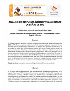
| dc.description.abstract | En el presente artículo, se analizó el proceso nociceptivo mediante señales de electroencefalografía (EEG) de 17 voluntarios, quienes recibieron un estímulo nociceptivo y durante la presencia de este, evaluaron el dolor percibido según la escala EVA. La señal de EEG se analiza en línea base y en el momento en que se tiene el valor de dolor más alto de la escala. Para el análisis, se hizo una previa remoción de artefactos presentes en los canales a trabajar mediante el uso de la transformada wavelet discreta. Una vez obtenida la señal filtrada, se hizo una descomposición en bandas de ondas cerebrales características theta, alfa, beta y gamma, usando filtros FIR; con el fin de ver la deferencia interhemisférica del valor RMS entre pares de canales complementarios. Se analizaron las diferencias interhemisféricas de línea base contra las de dolor máximo en cada una de las bandas encontrando que, en promedio, la variación más significativa se encuentra en onda theta y en los canales ubicados en regiones correspondientes a zona media del cerebro. | spa |
| dc.description.abstract | In this article, the nociceptive process was analyzed using electroencephalography signals (EEG) of 17 volunteers, who received a nociceptive stimulus and during its presence, evaluated the perceived pain according to the VAS scale. The EEG signal is analyzed at baseline and at the moment when the highest pain value on the scale is reached. For the analysis, a prior removal of artifacts present in the channels to work through the use of discrete wavelet transform. Once the filtered signal was obtained, a decomposition into characteristic theta, alpha, beta, and gamma brainwave bands, using FIR filters; with the final purpose to see the interhemispheric difference of the RMS value between pairs of complementary channels. I know analyzed the interhemispheric differences of baseline against those of maximum pain in each of the bands, finding that, on average, the most significant variation is found in wave theta and in channels located in regions corresponding to the midbrain. | eng |
| dc.format.extent | 91 páginas | spa |
| dc.format.mimetype | application/pdf | spa |
| dc.language.iso | spa | spa |
| dc.title | Análisis de respuesta nociceptiva mediante la señal de EEG | spa |
| dc.type | Documento de Conferencia | spa |
| dc.type.version | info:eu-repo/semantics/publishedVersion | spa |
| oaire.accessrights | http://purl.org/coar/access_right/c_abf2 | spa |
| oaire.version | http://purl.org/coar/version/c_970fb48d4fbd8a85 | spa |
| dc.contributor.datamanager | López López, Juan Manuel | |
| dc.identifier.url | https://catalogo.escuelaing.edu.co/cgi-bin/koha/opac-detail.pl?biblionumber=23082 | spa |
| dc.publisher.place | Cartagena, Colombia | spa |
| dc.relation.conferencedate | 21- 24 de septiembre 2021 | spa |
| dc.relation.conferenceplace | Cartagena, Colombia | spa |
| dc.relation.indexed | N/A | spa |
| dc.relation.ispartofconference | Encuentro Internacional de Educación en Ingeniería (EIEI) ACOFI 2021 | spa |
| dc.relation.references | Albu S, Meagher M W (2019) Divergent effects of conditioned pain modulation on subjective painand nociceptive‑related brain activity. Springer. | spa |
| dc.relation.references | Jensen M P, Hakimian S, Sherlin L H and Fregni F. (2008). New Insights Into Neuromodulatory Approaches for the Treatment of Pain. The Journal of Pain Vol 9. Pp 193-199. | spa |
| dc.relation.references | Lee M C, Mouraux A and Ianetti (2009) Characterizing the Cortical Activity through Which Pain Emerges from Nociception. The Journal of Neuroscience. Vol 29(24) pp.7909-7916. | spa |
| dc.relation.references | Ploner M, Sorg C, and Gross J. (2017) Brain Rhythms of Pain. Cellpress, Trends in cognitive sciences. Vol 21. | spa |
| dc.relation.references | Ripanpitak K, He S, Sönmezisik I, Morant T, Huang S Y, Yu W. (2021) Granger Causality-Based Pain Classification Using EEG Evoked by Electrical Stimulation Targeting Nociceptive A and C Fibers. IEEE Access. | spa |
| dc.relation.references | Sarnthein J and Jeanmonod D (2008). High thalamocortical theta coherence in patients with neurogenic pain. Elseiver NeuroImage, Vol. 39, pp. 446-462. | spa |
| dc.relation.references | Torta M E, Legrain V, Algoet M, Olivier E, Duque J and Mouraux A. (2013). Theta Burst Stimulation Applied over Primary Motor and Somatosensory Cortices Produces Analgesia Unrelated to the Changes in Nociceptive Event-Related Potentials. Plos One Vol 8. | spa |
| dc.relation.references | Valentini E, Betti V, Hu L and Aglioti S M (2019). Hypnotic modulation of pain perception and of brain activity triggered by nociceptive laser stimuli. Cortex Vol 49. | spa |
| dc.relation.references | Youssef AM, Macefield V, and Henderson L (2016). Cortical Influences on Brainstem Circuitry Responsible for Conditioned Pain Modulation in Humans. Human Brain Mapping, Vol 37, pp. 2630–2644 | spa |
| dc.relation.references | Zhang Z G, Hu L, Hung Y S, Mouraux A, and Iannetti G D (2012) Gamma-Band Oscillations in the Primary Somatosensory Cortex—A Direct and Obligatory Correlate of Subjective Pain Intensity. The Journal of Neuroscience. Vol 32(22) pp. 7429 –7438. | spa |
| dc.relation.references | Purves D, Augustine G, Fitzpatrick D, Hall W, LaMantia A. y White L. (1997) Neuroscience. 5ta edición. pp. 29 - 127. | spa |
| dc.rights.accessrights | info:eu-repo/semantics/openAccess | spa |
| dc.subject.armarc | Electroencefalografía | |
| dc.subject.armarc | Nocicepción | |
| dc.subject.armarc | cerebro | |
| dc.subject.proposal | Electroencefalografía | spa |
| dc.subject.proposal | Electroencephalography | eng |
| dc.subject.proposal | Nocicepción | spa |
| dc.subject.proposal | Nociception | eng |
| dc.subject.proposal | Cerebro | spa |
| dc.subject.proposal | Brain | eng |
| dc.type.coar | http://purl.org/coar/resource_type/c_18cp | spa |
| dc.type.content | Text | spa |
| dc.type.driver | info:eu-repo/semantics/article | spa |
| dc.type.redcol | http://purl.org/redcol/resource_type/ART | spa |
Ficheros en el ítem
Este ítem aparece en la(s) siguiente(s) colección(ones)
-
BA - Trabajos Dirigidos de Biomédica [227]
Trabajos del Pregrado de Ingeniería Biomédica de la Escuela Colombiana de Ingeniería Julio Garavito


